On this page
How to identify it
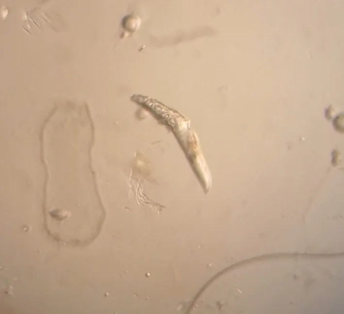
- Adult Demodex canis are cigar-shaped mites with four pairs of stumpy legs near the front of the body. Males reach about 250 micrometers (0.25 mm) long and females about 300 micrometers (0.3 mm). Veterinarians find them in deep skin scrapings.[1]
- Dogs can carry D. canis, the most common species, and the longer D. injai. A short form called D. cornei is also described, but whether it is a separate species is disputed.[1][3]
- Localized mange in dogs shows as one to four small patches of hair loss, redness and silvery scaling, often around the mouth, eyes and front legs.[1]
- Generalized mange covers large areas or whole body regions with hair loss, blackheads (comedones), follicular casts, bumps and pustules. Severe cases develop crusting and secondary bacterial infection, and the disease can also be confined to the feet.[1][3]
- Itching is usually not a main sign of demodectic mange in dogs unless the skin has a secondary bacterial infection.[1][3]
- D. injai turns up more often in terriers and makes the coat and skin excessively greasy.[3]
- In cats, D. cati looks like the dog mite but narrower and causes similar signs, often on the eyelids, around the eyes and on the head and neck. D. gatoi is smaller with a broad, blunt rear end, lives closer to the skin surface and causes intense itching, hair loss, scaling and crusts, mostly on the head, neck and elbows, with itching of the trunk also reported.[2][3]
Look-alikes
| Look-alike | How to tell them apart |
|---|---|
| Ear mites (Otodectes cynotis) | Ear mites are surface mites with long legs that live in the external ear canal. Demodex mites live in hair follicles, and only occasionally fill the ear canals and cause a waxy outer ear inflammation.[4][1][2] |
Where it lives
Demodex canis occurs on dogs around the world, and demodicosis is a common disease in small animal practice worldwide. The cat mite D. gatoi was long considered very regional and was mostly diagnosed in the Southeastern United States, but it has since been reported in cats in other parts of the world.[1][3]
Biology and life cycle
The whole life cycle takes place on the animal. Adults live mainly in the hair follicles, where females lay eggs; each egg hatches into a larva that molts through larval and nymph stages to the adult. The cycle takes at least three weeks.[1]
Puppies pick up the mites from their mother during the first days of nursing. Direct spread from dog to dog under natural conditions seems to be very rare. The mites do not survive well off the animal, and mites off the host are generally not thought to infect dogs.[1][3]
Demodex species are highly specific to their hosts: dog mites do not infest other kinds of animals.[1]
Disease develops in dogs that carry larger numbers of mites, probably because of an immune deficit. In young dogs a temporary immune change and a genetic basis seem to be involved; in older dogs the disease may go with an immune-suppressing illness or treatment. It is more common in purebred than mixed-breed dogs, and often a whole susceptible litter is affected.[1][3]
Suggested triggers include poor nutrition, heat cycles and giving birth, immune-suppressing drugs and chronic disease.[1]
In cats, demodicosis is usually tied to another disease, except for D. gatoi, which spreads between cats and can also affect otherwise healthy ones.[2][3]
Damage and signs
- Localized demodicosis is mostly seen in dogs under a year old and often clears without treatment. Patches may come and go for several months, but once they resolve they rarely return, and this form does not often progress to the generalized disease.[1]
- Generalized demodicosis is among the most severe canine skin diseases. It usually first appears in puppies 3 to 18 months old, and these juvenile cases have a good outlook with modern treatment. When it first appears in a dog older than five years it usually points to an underlying disease, such as cancer, that weakens the immune system, and the outlook is generally poor.[1]
- Left untreated, generalized disease can cover the skin with crusted, infected, bleeding lesions, mainly on the head and legs, after several months.[1]
- In cats, generalized disease from D. cati is rare and usually linked to an underlying illness such as diabetes, feline leukemia virus or feline immunodeficiency virus.[2]
Health risk
People carry their own species of Demodex, mainly in the follicles of the eyelashes and eyebrows, without harm, and cannot be infected with dog or cat Demodex mites.[1][2]
Control
Prevention
Dogs that have had generalized demodicosis, and their parents, should not be used for breeding, because the juvenile disease has a genetic basis.[1][3]
Pesticides
Isoxazoline drugs are now the drugs of choice for canine demodectic mange, and a moxidectin and imidacloprid combination carries a label claim for Demodex. The WAVD panel rates amitraz and macrocyclic lactones such as ivermectin as effective too, though WCVM describes amitraz dips and lime sulfur baths as largely obsolete for dogs. For cats, the strongest evidence is for lime sulfur dips and amitraz baths, and all cat treatments are used off-label.[1][2][3]
Ivermectin can cause severe, sometimes fatal nerve poisoning in dogs that carry a mutation of the ABCB1 gene (formerly called MDR1). This is seen most often in collies, Australian shepherds, Shetland and Old English sheepdogs and their crosses, but also in other breeds, and the WAVD panel recommends a gradual dose increase when starting ivermectin or moxidectin given by mouth, to identify sensitive dogs.[3]
Amitraz rinses should be done in a well-ventilated area by a handler wearing protective clothing, because breathing problems and other side effects have been reported in people.[3]
When to call a professional
Mange needs a veterinarian's diagnosis. Because Demodex mites are normal skin residents, finding them only means something alongside the pet's history and signs. Deep skin scrapings, taken while squeezing a fold of skin and sometimes from several sites, are the method of choice; plucked hairs, tape strips and skin biopsies can also help.[1][2][3]
Localized mange in dogs usually needs no treatment unless it fails to clear within a few months or spreads. Generalized disease is treated and checked clinically and microscopically every month, and the WAVD guidelines advise continuing treatment for four weeks after the second negative monthly scraping to lower the chance of relapse.[1][3]
When D. gatoi is found, every cat in contact with the affected one should be treated, not just the cat with signs.[2]
Common questions
- Can I catch demodectic mange from my dog or cat?
- No. People have their own Demodex species, mostly in eyelash and eyebrow follicles, and cannot be infected with dog or cat Demodex mites.[1][2]
- Will my puppy's mange spread to my other dogs?
- Unlikely. Puppies get the mites from their mother while nursing in the first days of life, and direct spread between dogs under natural conditions seems to be very rare. In cats the exception is D. gatoi, which is contagious, so all cats in contact are treated.[1][2][3]
- Does a small bald patch on my puppy need treatment?
- Often not. Localized demodicosis in dogs under a year old usually clears by itself; treatment is used if it does not clear within a few months or progresses to the generalized form.[1]
Sources
- University of Saskatchewan, Western College of Veterinary Medicine, “Demodex canis (Learn About Parasites, March 2021).” wcvm.usask.ca/learnaboutparasites/parasites/demodex-canis.php (accessed 2026-11-08)
- University of Saskatchewan, Western College of Veterinary Medicine, “Demodex species: cats (Learn About Parasites, March 2021).” wcvm.usask.ca/learnaboutparasites/parasites/demodex-species-cats.php (accessed 2026-11-08)
- Veterinary Dermatology (PDF hosted by the World Association for Veterinary Dermatology), “Diagnosis and treatment of demodicosis in dogs and cats: clinical consensus guidelines of the World Association for Veterinary Dermatology (R. S. Mueller and colleagues, Veterinary Dermatology 31: 4-e2, 2020).” wavd.org/wp-content/uploads/diagnosis-and-treatment-of-demodicosis-in-dogs-and-cats-mueller-et-al-2020-veterinary-dermatology.pdf (accessed 2026-11-08)
- University of Saskatchewan, Western College of Veterinary Medicine, “Otodectes cynotis (Learn About Parasites).” wcvm.usask.ca/learnaboutparasites/parasites/otodectes-cynotis.php (accessed 2026-11-08)
- iNaturalist, “Demodex canis taxon page (order and family).” www.inaturalist.org/taxa/262784 (accessed 2026-11-08)